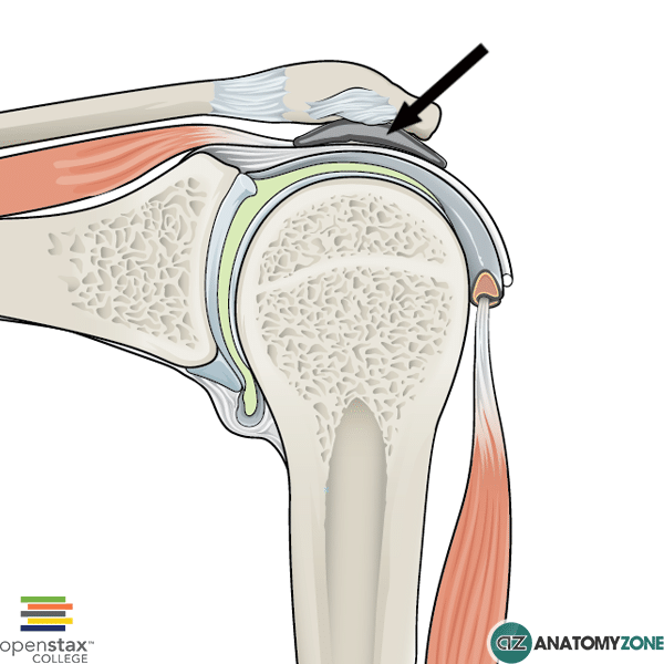
Subacromial Bursa
The structure indicated is the subacromial bursa.
The glenohumeral joint is a synovial ball and socket joint between the head of the humerus and the glenoid cavity of the scapula. It is a joint with great mobility, at the expense of joint stability.
Movements include: abduction, adduction, medial rotation, lateral rotation, flexion, extension, and circumduction.
The joint is stabilised by, surrounding musculature, tendons, ligaments and the bony processes of adjacent structures.
The synovial membrane lines the fibrous membrane of the joint capsule and it forms bursae as it passes through various openings in this membrane. Synovial bursae are fluid-filled (synovial fluid) sacs lined by synovial membrane, providing a cushion between bones, tendons and muscles, essentially lubricating the joint and allowing free, fluid movement.
There are several bursae associated with the shoulder joint.
The subacromial bursa, as the name suggests, is located below the acromion. In most individuals it communicates with the subdeltoid bursa to form the subacromial-subdeltoid bursa.
The subacromial bursa separates the superior surface of the supraspinatus tendon from the acromion, the coracoid, and the coraco-acromial ligament which lie above. The bursa is important in providing fluid motion of the supraspinatus tendon.
A common painful shoulder condition arises when this bursa becomes inflamed – subacromial bursitis, which is often part of impingement syndrome where the tendons of the rotator cuff muscles become inflamed as they pass through the subacromial space.